Scanning Electron Microscope (SEM)
Background: To characterize the nanoscale structure prepared by the alkaline oxidation to the copper substrate.
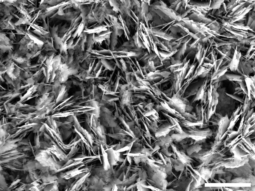
From the SEM image in the top view (Figure 1), I found that the alkaline oxidation to copper introduces a large amount of nanoscale flake-like structure, which has a random orientation. Also, the thickness and width of the flake-like structure are ~100nm and ~2μm. However, the SEM image in the top view cannot provide information on the height and inter-distance of the flake-like structure.
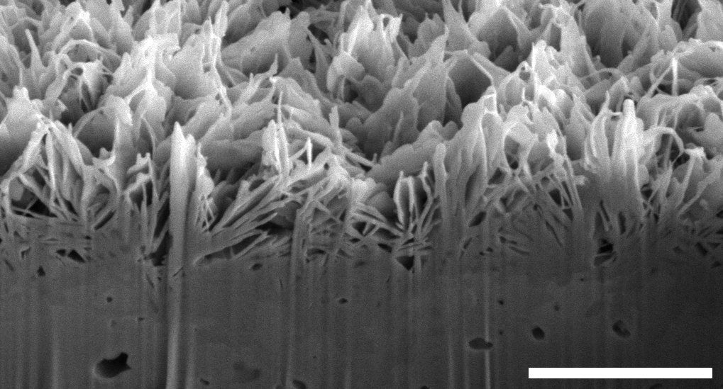
Therefore, I prepared the cross-section sample using the focus electron beam (FIB) technique to obtain comprehensive information. From the SEM image of the cross-section sample (Figure 2), I found that the nano-flake structure has a height and inter-distance of ~1μm and ~300nm. In addition, the nano-flake structure grows in various directions, instead of perpendicular to the substrate.